The History of Cancer: From Ancient Crabs to Cellular Code
Cancer feels like a modern disease, but it is as old as multicellular life. Dinosaur bones carry tumours. Egyptian mummies carry them too. What is new is our understanding of it. In about 3,600 years, humanity has moved from writing "there is no treatment" on a papyrus to reading the mutations of a single tumour and designing a drug, or even a vaccine, around them. This article follows that journey: what cancer is, how it was discovered, and what medicine uses against it today.
What is cancer?
Cancer is not a single disease but a family of more than one hundred different diseases that share one frightening feature: a group of the body's own cells stops obeying the rules that keep a living creature organised, and begins to grow, spread and survive for its own sake. To see why this is so serious, it helps to picture the body as a country of roughly thirty trillion citizens. Every cell has a job. Heart cells pump blood, nerve cells carry electrical signals, lung cells swap carbon dioxide for oxygen, liver cells clean the blood of chemicals, and white blood cells patrol for infection. The citizens constantly talk to one another through chemical messages that tell each cell when to grow, when to divide, when to rest and, when necessary, when to die. A cell that is badly damaged is supposed to destroy itself in a tidy, controlled way called apoptosis, or programmed cell death, so that it cannot pass its damage on. Cancer is what happens when a cell ignores these orders, and its descendants keep ignoring them.
The orders themselves are written in DNA, the long molecule inside every cell that holds our genes. A gene is a stretch of DNA that carries the recipe for one protein, and proteins do the actual work of the cell. Some genes act like the accelerator pedal of growth, and when they are damaged so that they are stuck in the on position they are called oncogenes (the healthy version is a proto-oncogene). Well-known examples are RAS, MYC and HER2. Other genes act like brakes, or like a mechanic who checks the engine, and these are called tumour suppressor genes. TP53, nicknamed the guardian of the genome, halts a damaged cell and forces it to repair itself or die, while BRCA1 and BRCA2 help to repair broken DNA. A permanent change in the DNA sequence is called a mutation, and mutations arise all the time: from random copying mistakes whenever a cell divides, from tobacco smoke and other chemical carcinogens, from ultraviolet light and ionising radiation, from infections such as human papillomavirus (HPV), hepatitis B and C and the stomach bacterium Helicobacter pylori, and occasionally from a faulty gene inherited from a parent.
Most mutations are harmless or are quickly repaired. Trouble begins when mutations strike the accelerators and the brakes themselves, and when several of them pile up in the same cell. Scientists call the damaging hits driver mutations and the harmless bystanders passenger mutations. There are also epigenetic changes, which switch genes on or off without altering the DNA letters, for example by attaching chemical tags (a process called methylation) to a gene. Cancer, in other words, is a disease of accumulated damage, which is the main reason the risk rises so steeply with age. The two figures below show the long road a single cell must travel before it becomes a dangerous tumour.
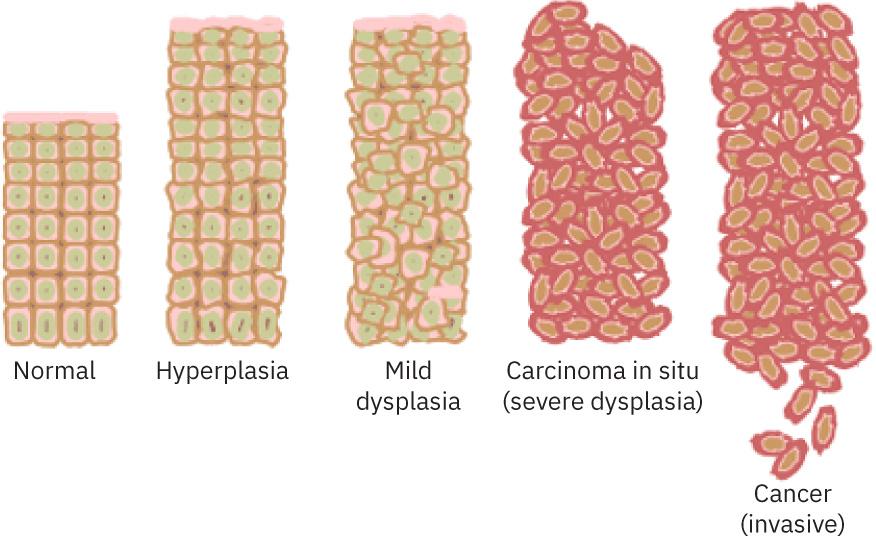
How a tissue changes on the way to cancer: from normal tissue, through hyperplasia (too many cells), mild dysplasia (cells that begin to look abnormal) and carcinoma in situ (severe dysplasia, still confined to its layer), to invasive cancer, in which cells break out of the tissue.
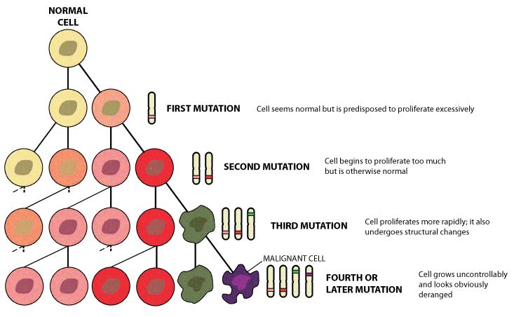
Cancer as accumulated damage. A cell with a first mutation still looks normal but is predisposed to multiply too much, and each further mutation in its descendants makes the clone grow faster and look more abnormal, until a fourth or later mutation produces an obviously malignant cell.
This slow build-up was put into a framework in 2000 by Douglas Hanahan and Robert Weinberg, who proposed that every cancer, whatever its organ, acquires the same set of abilities, which they called the hallmarks of cancer. The cell makes its own growth signals, ignores the signals that say stop, resists apoptosis, and divides without limit by switching on an enzyme called telomerase that rebuilds the protective caps (telomeres) at the ends of chromosomes. It persuades the body to build new blood vessels for it, a process called angiogenesis, and it learns to invade neighbouring tissue and travel to distant organs, which is called metastasis. Later versions of the framework added a reprogrammed appetite for energy and the ability to hide from the immune system, together with enabling features such as unstable DNA and chronic inflammation. A tumour is also not just a ball of identical cancer cells. It is surrounded by blood vessels, immune cells, connective tissue and signalling molecules that together form the tumour microenvironment, and the cancer actively bends this neighbourhood to its advantage.
Doctors name cancers after the cell they start from. Carcinomas begin in epithelial cells, the cells that line the skin, the airways, the gut and the glands, and they make up the large majority of cases, including lung, breast, prostate and colon cancer. Sarcomas begin in bone, muscle, fat and other connective tissue. Leukaemias begin in the blood-forming cells of the bone marrow, lymphomas in the lymph nodes and the wider lymphatic system, and myelomas in plasma cells, the cells that make antibodies. Tumours of the brain and spinal cord form a group of their own. The word cancer therefore tells us very little by itself, because a slow thyroid cancer that is cured by surgery and an aggressive pancreatic cancer have almost nothing in common except the name. A measurable feature of a tumour, such as a protein on its surface or a particular mutation, is called a biomarker, and modern treatment depends heavily on finding the right one.
The scale of the problem is large. Global estimates for 2022 (GLOBOCAN) counted roughly 20 million new cases and about 9.7 million deaths in that single year, which makes cancer one of the leading causes of death in the world.
How cancer harms and kills
A small group of abnormal cells may do no noticeable harm for years. The danger starts as the population grows and begins to behave like an invading society. First, it takes up space and destroys normal tissue. This matters most where there is no room to spare: the brain sits inside a rigid skull, so even a modest tumour can press on its surroundings and disturb movement, speech, vision or consciousness, while a tumour in the lung can block an airway or replace the working tissue that exchanges oxygen. Second, it steals resources. Cancer cells are hungry, and as a tumour outgrows its blood supply it releases chemical signals that make nearby vessels sprout new branches into it. This angiogenesis feeds the tumour with oxygen and glucose, which is why drugs such as bevacizumab, which cut off this supply, became a form of treatment. Third, it ignores boundaries. Normal cells stay in their own tissue, held in place by a thin sheet called the basement membrane, whereas cancer cells release enzymes that dissolve this barrier and push into the neighbouring tissue. A growth that stays put is called benign, and a growth that invades is called malignant, which is the true meaning of the word cancer.
| Benign tumour | Malignant tumour | |
| Invades neighbouring tissue | No, it stays local | Yes |
| Spreads to distant organs | No | Yes, by metastasis |
| Usual danger | Low, unless it sits in a critical place such as the skull | Can be life-threatening |
The most dangerous ability of a malignant tumour is metastasis, the spread of cancer to distant organs. Some cells break away from the original, or primary, tumour, enter a blood vessel or a lymph vessel, survive the journey and the attack of the immune system, settle in another organ and begin to multiply there. Lung cancer commonly spreads to the brain, liver, bones and adrenal glands, and breast cancer to bone, lung and liver. A lung cancer that has reached the liver is still called metastatic lung cancer, not liver cancer, because its cells keep the identity and the mutations of their origin. Metastasis is the reason that most cancer deaths are not caused by the first tumour. A tumour found early and removed is often cured, while a cancer that has already seeded many organs is much harder to clear.
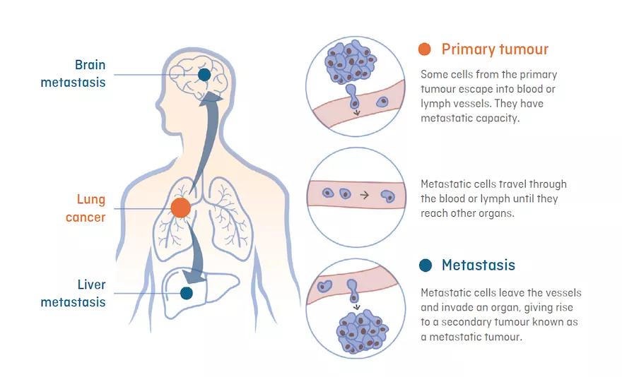
How metastasis happens, using lung cancer as an example. Cells escape from the primary tumour into blood or lymph vessels, travel to other organs, leave the vessels and grow into a secondary tumour, here in the brain and the liver.
So how does cancer actually kill? Rarely because there are simply too many cancer cells, but because the cells interfere with the organs that keep us alive. Extensive cancer in the liver destroys its ability to clear toxins and make essential proteins, and the result is liver failure. Cancer in or spreading to the lungs blocks airways or damages the tissue that takes up oxygen, and the result is respiratory failure. Tumours in or near the brain can cause swelling, seizures and loss of vital functions. Leukaemias crowd out the normal blood-cell factories of the bone marrow, leaving the patient with severe anaemia, bleeding and infections the body cannot fight. Many people with advanced disease also develop cachexia, a wasting syndrome in which the tumour and the body's own inflammatory response rewire metabolism so that muscle and fat melt away even when the person is eating. Infections, blood clots and organ failure often combine in the final stage, which is why the cause of death in cancer is usually a failing organ rather than the tumour itself.
Why the immune system does not simply destroy cancer
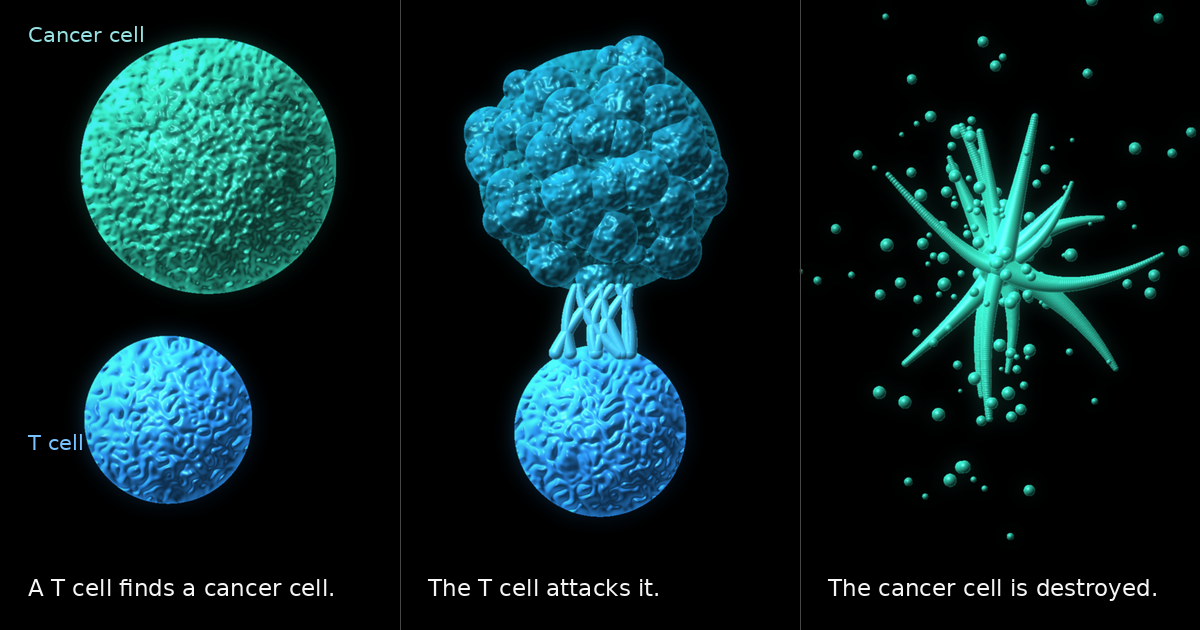
One of the great puzzles of the field is that the body already has an army for exactly this job. T cells and natural killer cells patrol the tissues, and they can recognise abnormal proteins, called neoantigens, that appear on the surface of mutated cells. This watch is known as immune surveillance, and it probably removes countless early cancers during a lifetime. But cancer is an evolutionary process. Among billions of dividing cells, the ones that happen to hide from the immune system or to disable it survive and multiply, while the rest are killed, a selection known as immunoediting. Tumours hide by switching off the molecules that display their abnormal proteins, they release signals that calm and exhaust immune cells, they recruit helper cells that suppress attack, and, most cleverly, they exploit immune checkpoints. A checkpoint is a brake that normally stops T cells from attacking healthy tissue, and the PD-L1 protein on a tumour cell can bind the PD-1 protein on a T cell and tell it to stand down. This explains why cancers grow even in people with a perfectly working immune system, and it is the weakness that modern immunotherapy attacks, as described later in this article.
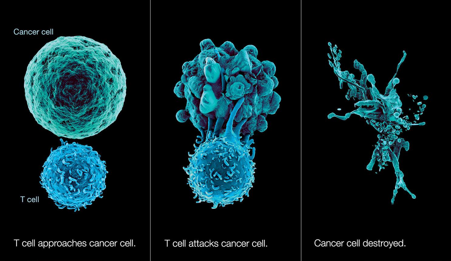
A T cell, one of the immune system's main killer cells, approaches a cancer cell, attacks it and destroys it. Immunotherapy aims to help T cells do this against cancers that have learned to hide from them.
How cancer was discovered: a timeline
Antiquity: naming the crab
The oldest known description is in the Edwin Smith Papyrus, an Egyptian surgical text copied around 1600 BCE and believed to draw on much older material. It describes bulging tumours of the breast, and the physician's verdict on treatment is blunt: there is none. Fossil and mummy evidence also shows bone tumours in ancient humans and in animals.
The Greek physician Hippocrates (c. 460 to 370 BCE) used the words karkinos and karkinoma, meaning crab, likely because a hard tumour with swollen veins radiating outward resembled a crab. The Roman writer Celsus translated this as cancer. Galen (2nd century CE) later used the term oncos, meaning swelling, which survives in the word oncology. Galen also held that cancer came from an excess of black bile, a humoral theory that dominated Western medicine for about 1,400 years.
The anatomical age (16th to 18th century)
Dissection began to undermine the humoral theory. In 1761 Giovanni Battista Morgagni of Padua linked disease to organ pathology through systematic autopsies, founding modern pathological anatomy. In 1775 the London surgeon Percivall Pott described scrotal cancer in chimney sweeps, the first clear link between an occupational exposure (soot) and cancer. It was the beginning of cancer epidemiology and of the idea of a carcinogen.
The cellular age (19th century)
The microscope changed everything. In 1838 Johannes Muller showed that tumours are made of cells. Around 1845 to 1847, John Hughes Bennett and Rudolf Virchow independently described leukaemia, a cancer of the blood. Virchow's principle, omnis cellula e cellula (every cell comes from a cell), led to the view that cancer cells arise from normal cells that have gone wrong.
Surgery advanced alongside anaesthesia (1846) and antisepsis. In 1894 William Halsted at Johns Hopkins performed the first radical mastectomy, removing breast, chest muscles and lymph nodes on the theory that cancer spreads outward in a contiguous manner. It dominated breast surgery for nearly a century, until trials in the 1970s and 1980s showed that smaller operations combined with radiotherapy worked as well.
The age of radiation and hormones (1895 to 1940)
In 1895 Wilhelm Rontgen discovered X-rays, and within a few years they were used to treat tumours. In 1898 Marie and Pierre Curie discovered polonium and radium, opening the era of radiotherapy. In 1896 the Scottish surgeon George Beatson showed that removing the ovaries could shrink advanced breast cancer, the first sign that cancer can depend on hormones.
On the theory side, Theodor Boveri proposed in 1914 that cancer arises from abnormal chromosomes. In 1915 Katsusaburo Yamagiwa and Koichi Ichikawa produced skin cancer in rabbits by painting coal tar on their ears, proving experimentally that chemicals cause cancer. In 1911 Peyton Rous showed that a virus could transmit a chicken sarcoma, an idea that was laughed at for decades before he won the Nobel Prize in 1966.
The chemotherapy revolution (1940s to 1970s)
The first chemotherapy came from a war. After the 1943 Bari harbour incident, doctors noticed that survivors exposed to mustard gas had very low white blood cell counts. Louis Goodman and Alfred Gilman used this lesson to treat lymphoma with nitrogen mustard, the first cancer chemotherapy.
In 1947 and 1948 Sidney Farber in Boston gave aminopterin, a folate antagonist, to children with acute lymphoblastic leukaemia and obtained temporary remissions. Min Chiu Li later cured choriocarcinoma with methotrexate in the 1950s. Combination chemotherapy, pioneered by Emil Freireich, Emil Frei and others, produced cures in childhood leukaemia and Hodgkin lymphoma in the 1960s.
At the same time, the link with lifestyle was established. In 1950 Richard Doll and Austin Bradford Hill, and independently Ernst Wynder and Evarts Graham, showed that smoking causes lung cancer. George Papanicolaou developed the Pap smear, and cervical cancer screening started to prevent cancer in the 1940s and after. In 1971 the United States passed the National Cancer Act, launching the so-called war on cancer.
The molecular age (1970s to 2000s)
The 1970s and 1980s brought the real explanation. In 1976 J. Michael Bishop and Harold Varmus showed that the cancer-causing gene of the Rous sarcoma virus has a normal counterpart in healthy cells: the first proto-oncogene (src). In 1971 Alfred Knudson proposed the two-hit hypothesis for retinoblastoma, which led to the discovery of tumour suppressor genes. In 1979 the p53 protein was found, and TP53 later turned out to be the most frequently mutated gene in human cancer. Bert Vogelstein and colleagues showed in 1990 that colorectal cancer arises through a stepwise accumulation of mutations.
Chromosomes gave another key. In 1960 Peter Nowell and David Hungerford found the Philadelphia chromosome in chronic myeloid leukaemia. Janet Rowley showed in 1973 that it is a translocation between chromosomes 9 and 22, which creates the fusion gene BCR-ABL. The cause of a cancer was, for the first time, a single identifiable molecular error. In the 1990s, BRCA1 (1994) and BRCA2 (1995) were identified, making genetic testing for hereditary breast and ovarian cancer possible.
The Human Genome Project (completed 2003) and the sequencing technologies that followed made it possible to read whole tumour genomes. Harald zur Hausen's work on HPV and cervical cancer, rewarded with the 2008 Nobel Prize, led to HPV vaccines from 2006, one of the first vaccines that prevents a cancer.
Strange and surprising stories from cancer science
Cancer science is full of stories that sound like fiction. In 1951 doctors at Johns Hopkins Hospital took a sample from the cervical tumour of Henrietta Lacks, a young mother who died of the disease that year, and passed it to a laboratory without asking her or her family. The cells, named HeLa after her, became the first human cells that would divide indefinitely in a dish, and they have since been used in thousands of studies, including work on the polio vaccine and on HPV, the virus that we now know caused her cancer. Her family learned of all this only decades later, and the case became a landmark in the debate about consent in medical research. Another forgotten pioneer is William Coley, a New York surgeon who in the 1890s injected bacteria into tumours after noticing that some cancer patients improved following a severe infection. His mixtures, called Coley's toxins, were unreliable and were pushed aside by radiotherapy and chemotherapy, but he is now often called the father of cancer immunotherapy, because we understand today that the immune system was doing the work.
Cancer is almost never contagious, but there are remarkable exceptions in animals. Tasmanian devils suffer from devil facial tumour disease, first noticed in the 1990s, in which the cancer cells themselves pass from animal to animal when they bite each other, so that the tumour behaves like a parasite and has pushed the species towards extinction in the wild. Dogs carry an even older example, a sexually transmitted tumour thought to have started from a single animal several thousand years ago and to have spread around the world ever since, and similar contagious cancers have been found in clams and mussels. A different puzzle is known as Peto's paradox, after the epidemiologist Richard Peto. An elephant or a whale has hundreds or thousands of times more cells than a mouse and lives much longer, so it should have far more chances to collect cancer-causing mutations, yet these giants do not die of cancer more often than we do. Studies of elephants found about twenty copies of the TP53 guardian gene, where humans have one, and their cells destroy themselves very readily when DNA is damaged, which may be one way they stay protected. Naked mole-rats, which live for decades, appear to resist cancer partly through a thick, sugar-based gel between their cells, and researchers study these animals hoping to copy their tricks.
Some discoveries turned into everyday tools. In the 1920s the German biochemist Otto Warburg noticed that tumours burn sugar in an odd, wasteful way, fermenting glucose into lactic acid even when plenty of oxygen is available, a quirk now called the Warburg effect. A century later it is the principle behind PET scans, in which a slightly radioactive form of sugar is injected and the greedy tumours light up on the image. Cancer research has also reshaped other fields. The study of tumour viruses led to the discovery of oncogenes, the sequencing of tumour genomes helped to create modern genetics, and the HPV vaccine, the first widely used vaccine that prevents a cancer, is already cutting cervical cancer sharply in countries with high uptake. The history of cancer is not only a story of defeat, but also of surprising turns.
How cancer is treated today
Modern oncology rests on three older pillars, surgery, radiotherapy and chemotherapy, and several newer ones: hormone therapy, targeted therapy, immunotherapy and cell and gene therapy. Treatment is now chosen by the tumour's biology as much as by its location, an approach called precision oncology.
Surgery and radiotherapy
Surgery still cures most solid tumours that are caught early. Radiotherapy uses ionising radiation to damage DNA in cancer cells. Modern forms include intensity-modulated radiotherapy (IMRT), stereotactic body radiotherapy (SBRT), proton beam therapy and brachytherapy, which place the dose more precisely and spare healthy tissue.
Chemotherapy in detail
Chemotherapy, often shortened to chemo, means treatment with drugs that kill cells, and the word usually refers to cytotoxic drugs (cyto means cell, toxic means poisonous) that attack cells while they are dividing. The idea rests on a simple observation: a cancer cell is, above all, a cell that divides too much, so a poison that stops cell division should harm the cancer more than the rest of the body. Every dividing cell must pass through a cycle of stages: a growth phase (G1), a phase in which it copies all of its DNA (S phase), a second preparation phase (G2) and finally mitosis (M phase), when the chromosomes are pulled apart and the cell splits in two. Cells that are not dividing rest in a quiet state called G0. Most chemotherapy drugs interfere with one of these steps, either by damaging the DNA, by blocking the building blocks needed to copy it, or by wrecking the machinery that pulls the chromosomes apart. A damaged cell with working brakes would normally stop and repair itself, but cancer cells have usually lost guardians such as p53, so they carry on dividing into trouble and finally die, often by apoptosis.
Chemotherapy is not truly selective. It cannot tell a cancer cell from a healthy cell that is also dividing quickly, and the body has several such tissues: the bone marrow that makes blood cells, the lining of the mouth and gut, and the hair follicles. This is the reason for the best known side effects, such as low blood counts, mouth ulcers, diarrhoea and hair loss, and it explains why every dose is a compromise between killing the tumour and sparing the patient. Doses are usually calculated from body surface area and given in cycles that last a few weeks, followed by a rest period so that healthy tissues can recover. A guiding idea, proposed by Howard Skipper in the 1960s as the log-kill hypothesis, is that a dose of drug kills a fixed fraction of the cancer cells rather than a fixed number, so that many cycles are needed to push a large tumour down towards zero.
The drug families are best understood by what they attack. Alkylating agents, which descend directly from the nitrogen mustard of the 1940s, attach chemical groups to DNA and bind its two strands together, so the strands can no longer separate to be copied. The platinum drugs, cisplatin, carboplatin and oxaliplatin, work in a similar way. Cisplatin was found by accident in the 1960s, when the scientist Barnett Rosenberg noticed that platinum electrodes in a bacterial culture stopped the bacteria from dividing, and after its approval in 1978 it turned testicular cancer from a usually fatal disease into one that is cured in the great majority of patients. Antimetabolites such as methotrexate, 5-fluorouracil and gemcitabine are impostors: they look like the building blocks of DNA and RNA, or like the vitamins needed to make them, and when the cell uses them by mistake the copying process stalls. Anthracyclines such as doxorubicin slide between the rungs of the DNA and poison an enzyme called topoisomerase II, which normally relieves the tension in coiled DNA, and other drugs such as irinotecan and etoposide block topoisomerases too. Finally, the mitotic inhibitors attack the microtubules, the protein threads that act as ropes pulling chromosomes apart. The taxanes, first isolated from the bark of the Pacific yew, freeze the ropes in place, while the vinca alkaloids, taken from the Madagascar periwinkle, stop them from forming at all.
| Drug family | Examples | What it does | Typical side effect |
| Alkylating agents | cyclophosphamide, temozolomide, melphalan | Bind DNA and cross-link its strands | Low blood counts, rarely a later leukaemia |
| Platinum compounds | cisplatin, carboplatin, oxaliplatin | Cross-link DNA | Nerve damage, kidney and hearing damage (cisplatin) |
| Antimetabolites | methotrexate, 5-fluorouracil, gemcitabine | Mimic DNA building blocks and stall copying | Mouth sores, diarrhoea, low blood counts |
| Anthracyclines | doxorubicin, epirubicin | Insert into DNA and poison topoisomerase II | Heart muscle damage at high total doses |
| Topoisomerase inhibitors | irinotecan, etoposide | Stop DNA from untangling during copying | Diarrhoea (irinotecan), low blood counts |
| Taxanes | paclitaxel, docetaxel | Freeze microtubules so the cell cannot divide | Nerve damage, hair loss |
| Vinca alkaloids | vincristine, vinblastine | Stop microtubules from forming | Nerve damage |
Single drugs rarely cure, so chemotherapy is almost always given as a combination of drugs with different targets and different side effects, which attacks the tumour from several sides and makes it harder for resistant cells to survive. Famous combinations are known by their initials, such as ABVD for Hodgkin lymphoma, R-CHOP for non-Hodgkin lymphoma, BEP for testicular cancer and FOLFOX for colon cancer. The aim of treatment varies. It can be curative, as in childhood acute lymphoblastic leukaemia, where about nine in ten children in high-income countries are now cured, and in Hodgkin lymphoma and testicular cancer. It can be adjuvant, given after surgery to destroy cells that may have escaped and could cause metastases, or neoadjuvant, given before surgery or radiotherapy to shrink a tumour. And it can be palliative, used to control symptoms and extend life when cure is not possible. Most drugs are given into a vein, often through a port or a thin tube placed under the skin, but some are tablets, for example capecitabine, and others are injected into the fluid around the spinal cord to reach the brain.
The side effects are the part of chemotherapy that people fear most, and also the part where medicine has improved most. Nausea and vomiting, once almost unavoidable, are now largely prevented by drugs such as ondansetron, which blocks serotonin signals in the gut and the brain, together with aprepitant and steroids. A fall in white blood cells, called neutropenia, is dangerous because even a mild infection can become life-threatening, so a fever during chemotherapy is treated as an emergency, and growth factors such as filgrastim can help the marrow recover. Anaemia and a low platelet count cause tiredness and bleeding. Some drugs have signature harms of their own: anthracyclines can weaken the heart muscle after a high cumulative dose, cisplatin can injure the kidneys and the hearing, and taxanes, vincristine and platinum drugs can damage nerves and cause numbness or tingling in the hands and feet. Hair loss can sometimes be reduced by cooling the scalp, and many people describe fatigue and difficulty concentrating, sometimes called chemo brain. Some drugs affect fertility, so options such as freezing eggs or sperm are discussed beforehand, and a few, especially alkylating agents, slightly raise the long-term risk of a second cancer such as leukaemia. A modern advance is testing the patient's genes before treatment: people who carry certain changes in the DPYD gene break down 5-fluorouracil poorly and can suffer severe toxicity, so their dose is lowered.
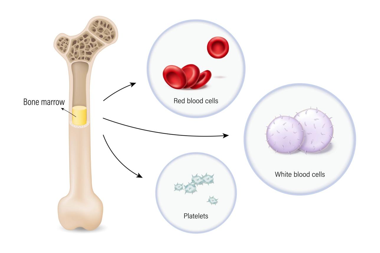
Blood cells are made in the bone marrow. Because chemotherapy damages fast-dividing tissue, the marrow is one of its main casualties, which is why patients can develop anaemia (too few red cells), infections (too few white cells) and bleeding (too few platelets).
Chemotherapy fails when cancer cells find a way around it. Some cells pump the drug out through molecular pumps in the cell membrane, notably one called P-glycoprotein, some repair the DNA damage faster, some mutate or multiply the target of the drug, and some sleep in the resting G0 state where most drugs cannot touch them. Tumours are also made of many genetically different clones, so treatment kills the sensitive ones and lets the resistant ones take over, which is a Darwinian process in miniature. Poorly supplied regions in the middle of a tumour may receive too little drug at all. For these reasons chemotherapy is now rarely used alone for many cancers. It is combined with immunotherapy, as in the first treatment of some lung cancers, with targeted drugs and with radiotherapy, and newer designs deliver the poison more precisely, such as liposomes that wrap the drug in a fatty bubble and the antibody-drug conjugates described below, which carry a chemotherapy payload straight to cells that display a certain protein. Chemotherapy is therefore not outdated, but is being reshaped from a blunt instrument into a more precise one.
Hormone (endocrine) therapy
Breast and prostate cancers often depend on hormones. Tamoxifen blocks the oestrogen receptor, aromatase inhibitors (letrozole, anastrozole) cut oestrogen production, and androgen deprivation therapy with drugs such as abiraterone and enzalutamide treats prostate cancer. This is the direct descendant of Beatson's 1896 observation.
Targeted therapy
Targeted drugs attack a specific molecular weakness. The landmark was imatinib (Gleevec, approved 2001), which blocks the BCR-ABL protein in chronic myeloid leukaemia and turned a fatal disease into a manageable one. Others include trastuzumab (HER2-positive breast cancer), EGFR inhibitors such as osimertinib in lung cancer, ALK inhibitors, BRAF and MEK inhibitors in melanoma, PARP inhibitors (olaparib) in BRCA-mutated cancers, CDK4/6 inhibitors (palbociclib) in breast cancer, and antiangiogenic agents such as bevacizumab.
Antibody-drug conjugates (ADCs) combine a targeting antibody with a potent chemotherapy payload, for example trastuzumab deruxtecan and sacituzumab govitecan. They act like guided missiles.
Immunotherapy
The immune system can recognise cancer, but tumours learn to hide. Immune checkpoint inhibitors release the brakes on T cells. James Allison (CTLA-4) and Tasuku Honjo (PD-1) shared the 2018 Nobel Prize in Physiology or Medicine for this work. The first checkpoint inhibitor, ipilimumab, was approved in 2011. Pembrolizumab and nivolumab (anti-PD-1), and atezolizumab (anti-PD-L1), followed. The Cancer Research Institute counted 156 FDA approvals for cancer immunotherapy from 2011 to the end of 2025, and subcutaneous versions of pembrolizumab, atezolizumab and nivolumab now reduce the time patients spend in the clinic.
CAR-T cell therapy takes a patient's own T cells, engineers them with a chimeric antigen receptor and returns them to attack tumour cells. The first CAR-T product, tisagenlecleucel, was approved in 2017 for childhood leukaemia. Bispecific antibodies, such as teclistamab in myeloma, bring T cells and tumour cells together physically. Cancer vaccines and oncolytic viruses, which are viruses engineered to infect and destroy tumour cells, complete the picture.
What is new in 2026
The past year has been busy. Reports from the FDA and the trade press describe several developments:
In August 2026 the FDA granted accelerated approval to vusolimogene oderparepvec (Tudriqev), an engineered oncolytic virus used with nivolumab for advanced melanoma that progressed after PD-1 therapy.
In the same month, the companies behind the INTerpath-001 trial announced positive phase 3 results for an individualised mRNA neoantigen therapy in resected melanoma, described as the first positive phase 3 trial for this type of treatment. Results come from company announcements, so independent peer-reviewed detail is still awaited.
In May 2026 the PROTAC drug vepdegestrant was approved for a subset of advanced breast cancer. PROTACs are a new class that does not just block a protein but tags it for destruction by the cell's own proteasome (targeted protein degradation).
In October 2026 the FDA gave accelerated approval to dostarlimab for locally advanced rectal cancer with mismatch repair deficiency (MSI-H/dMMR). Earlier in the year the device Optune Pax, which delivers tumour treating fields, was approved for locally advanced pancreatic cancer alongside chemotherapy.
A general lesson is that therapies are moving earlier in the disease, and that combinations of chemotherapy, targeted drugs and immunotherapy are becoming common. Approval status changes quickly, so readers should check current regulators' lists.
Screening and prevention
The cheapest progress comes from prevention. Tobacco control, HPV and hepatitis B vaccination, sun protection, and screening with mammography, colonoscopy, low-dose CT for high-risk smokers and cervical cytology or HPV testing save many lives. Liquid biopsy, which detects circulating tumour DNA in blood, and multi-cancer early detection tests are active research areas, but their real-world value is still under evaluation.
Challenges that remain
Drug resistance is common: tumours are genetically diverse, and treatment selects the cells that survive (clonal evolution). Immunotherapy works only in a subset of patients and can cause immune-related side effects. New drugs are very expensive, and access is unequal between rich and poor countries. Some cancers, such as pancreatic cancer and glioblastoma, are still difficult to treat. Research continues into artificial intelligence for pathology and drug discovery, personalised neoantigen vaccines, and better early detection.
Conclusion
The story of cancer is the story of how medicine learned to look closer: first at the body, then at the organ, then at the cell, and now at the gene and the molecule. The crab of Hippocrates has become a map of mutations, and for many patients, cancer has become a chronic illness or a cured one. Yet the disease is still among the leading causes of death worldwide, and the history teaches humility as well as hope.
Note for readers: this article is for education and does not replace medical advice. Treatment decisions belong with a qualified oncologist.
Further reading
Siddhartha Mukherjee, The Emperor of All Maladies: A Biography of Cancer (2010).
Douglas Hanahan and Robert Weinberg, Hallmarks of Cancer, Cell (2000) and Hallmarks of Cancer: The Next Generation, Cell (2011).
Cancer Research Institute, Cancer Immunotherapy Insights and Impact report (2026).
U.S. Food and Drug Administration, Oncology (Cancer) Approval Notifications.
F. Bray et al., Global cancer statistics 2022: GLOBOCAN estimates, CA: A Cancer Journal for Clinicians (2024).